Did you know there is the existence of ‘extra’ bones in the feet that can potentially and directly cause pain in the feet? There are several of these, and they can all play a role in foot pain. The human foot has the potential to grow small round bones that are not a part of normal human anatomy. While the location of these bones tends to stay consistent from person to person, not every human has them, and therefore they are considered ‘extra’.
I would like to start with a bone called the os naviculare. This small round bone is often found on the inner side of the middle of the foot (the same side as the big toe, but further back towards the ankle). This bone is found on the side of the navicular bone, and sometimes is actually fused into the navicular. The bone can cause pain by simply being prominent on the side of the foot, irritated by the side of the shoe. It can also cause pain by irritating an important tendon attached to the side of the navicular, called the posterior tibial tendon. The os naviculare bone can rub into the tendon during foot motion, and create inflammation and tendon degeneration over time. Sometimes, this bone can even form in between the fibers of the posterior tibial tendon, leading to tendon irritation from within.
Like all of these extra bones, treatment typically involves padding, wider shoes, or surgical removal of the bone itself.
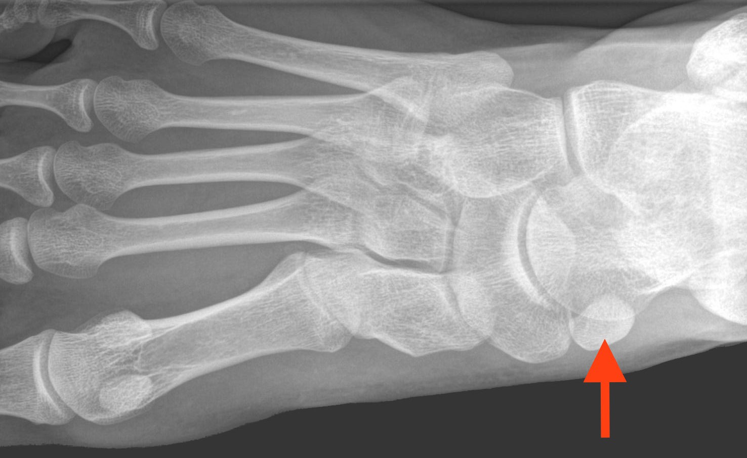
The next extra bone in the foot that is a source of discomfort is located on the other side of the os naviculare, which I discussed last post. The os peroneum is located on the outer side of the foot, near the middle of the foot’s length. This round to oval shaped bone is found either under or within the dual tendons of the peroneal muscles. These tendons help the peroneal muscles pull the foot outward, and they also act as a stabilizer for the foot when walking on uneven terrain. It is not uncommon for one or both of these tendons to become injured and inflamed. The os peroneum can also contribute to this injury by irritating the overlying tendon. Tight shoes can further this irritation, as can direct impact against the side of the foot.
Interestingly, this bone is sometimes mistaken for a fracture fragment in emergency rooms, especially if it is a little more jagged in appearance and lies close to the base of the 5th metatarsal bone, a long bone in the side of the foot that has a part which flares out into the side of the foot.
Again, like all of these extra bones, treatment typically involves padding, wider shoes, or surgical removal of the bone itself, along with a likely repair of the tendon near the bone.
The next extra bone in the foot that causes pain is located behind the ankle. This bone, also called os trigonum, is located behind the ankle just off the bone that forms the foot portion of the ankle joint, the talus. This extra bone is likely part of the talus that never fused into it’s main body, instead the bone is connected by a fibrous tissue band. This bone can be very small, or it can be somewhat large.
The os trigonum typically sits quiet for years, causing no pain. However, injuries such as an ankle sprain, or chronic activities that point the toe downward like ballet or soccer can cause the bone to become injured and inflamed. A nutcracker-like effect is created on the bone during these scenarios as the heel crunches the os trigonum into the ankle. This leads to bone bruising, injury of the fibrous tissue connecting the os trigonum to the talus, or sometimes even fracturing of the bone itself.
The pain felt with this condition can include a deep aching in the back of the ankle when the foot bends downward, pain with direct pressure to the back of the ankle, or swelling behind the ankle.
Treatment is a little different than the last two bones discussed. It involves immobilization and anti-inflammatory medications more to reduce the inflammation, as padding and shoe size change does not help much. A carefully placed steroid injection may also be of some benefit. If these measures do not help, surgery can be performed to remove the non-vital bone.
The final extra bone in the foot that causes pain that I will cover is located underneath the big toe. The big toe contains two bones (called phalanx bones). The other toes each have three, but the big toe is different for an anatomic reason well beyond the point of this post topic. These two bones form a joint where they come together, which is seen externally as the ‘knuckle’ in the big toe. For most people, that is it. However, some humans have formed an extra bone underneath this joint. This extra bone is often referred to as a ‘sesamoid’, but it should not be confused with the normal two sesamoid bones that everyone has underneath the big toe joint at the base of the big toe.
This extra bone is not always felt, or more specifically it does not always cause tissue irritation that leads to foot pain. Many people happily live with this bone and never experience any issues. However, this bone can potentially irritate the tendon that allows the big toe to bend downward if it is too prominent where the tendon rests under toe. It can also limited bending motion by directly contributing to or aggravating joint arthritis on the undersurface of the joint in the big toe. This bone is also implicated in diabetic skin wounds that form under the big toe, as the prominent bone often creates excessive pressure on the fragile skin below, leading to a pressure wound.
Treatment centers around accommodating the undersurface of the big toe with padding, an insert that is stiffened under this area to decrease the bending motion of the toe, or sometimes a steroid injection to reduce inflammation. Stiff soled shoes may also help. If these measures do not alleviate the problem, the bone can be simply removed through a small incision in the bottom of the toe.
